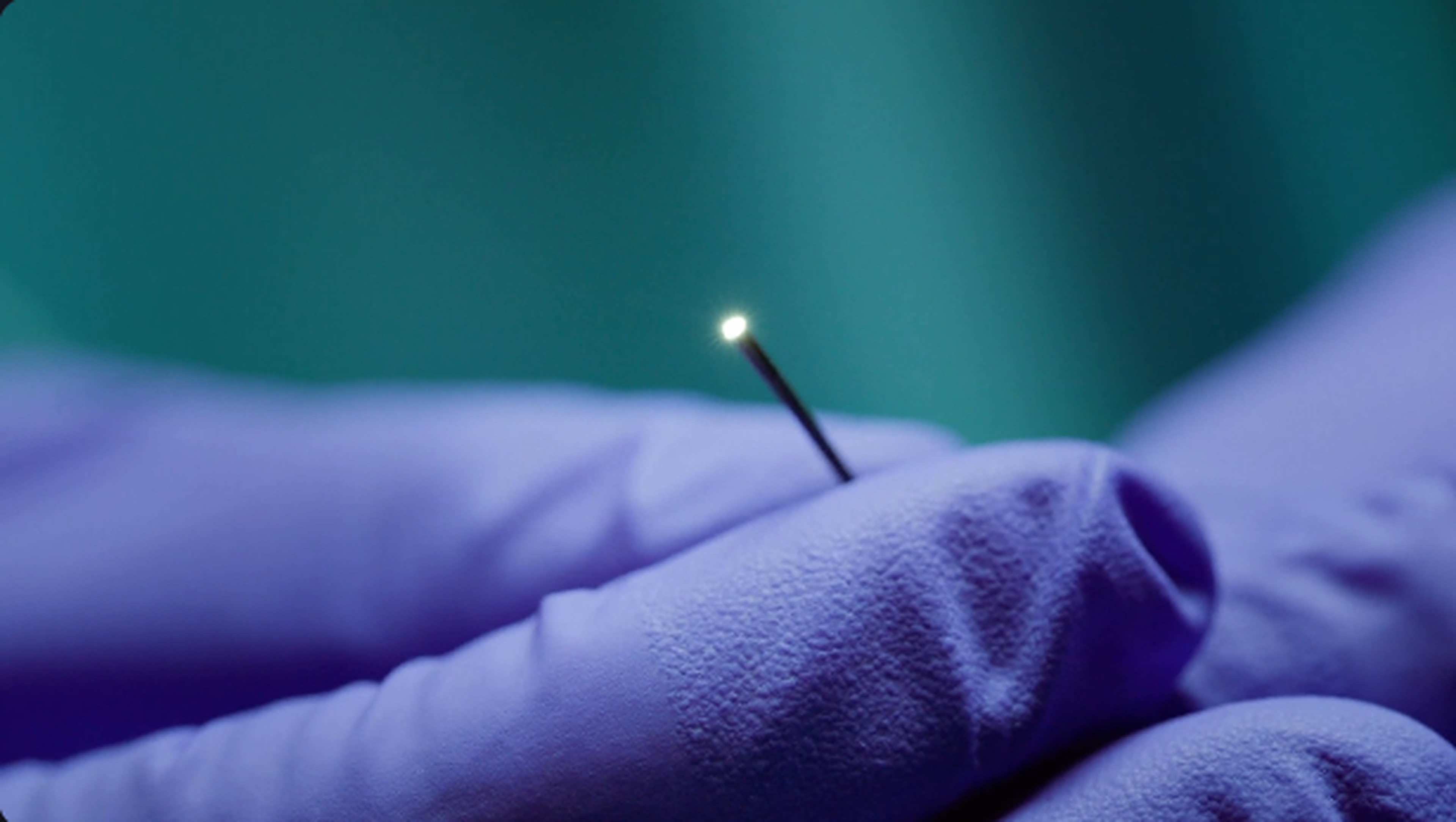
Vena Medical today announced that Health Canada has issued a Medical Device Licence for the Vena MicroAngioscope System™, authorizing its use to visualize the interior of blood vessels in the peripheral, coronary and neurovasculature. The first intravascular imaging device with a licensed neurovascular indication worldwide, is less than a millimeter in diameter and provides physicians with real‑time, full‑color, direct visualization from inside the blood vessel to complement existing imaging modalities, such as X-ray. Physicians are able to use this tool to quite literally see inside the blood vessel. This provides real-time feedback to help with decision-making as they treat their patients.
As of September 2025, the MicroAngioscope System has been used in over 30 commercial cases across Canada, including procedures performed at The Ottawa Hospital, Toronto Western Hospital, University of Alberta Hospital, and Royal University Hospital.
The MicroAngioscope System is available for sale and clinical use in Canada. While the Vena MicroAngioscope has been designated as a FDA Breakthrough Device, it has not been cleared or approved by the U.S. Food and Drug Administration.
Spotlight at PIKES 2025
Vena Medical will sponsor and present at PIKES 2025 in Toronto this week (September 25–27, Hotel X). Attendees can experience hands‑on demos of the MicroAngioscope System at the Vena booth and hear the clinical team during multiple sessions on direct intravascular visualization. To schedule a meeting at PIKES, please use the Contact Us form on Vena’s website www.venamed.ca.
Executive & Clinical Quotes
“Health Canada’s license opens a new era for real-time intravascular imaging throughout the entire vascular system, including the brain,” said Adam Karamath, Senior Director of Commercial Operations, Vena Medical. “With more than 30 successful commercial cases, Vena is bringing this technology into Canada first, where clinical adoption is beginning to show benefits already.
“Direct visualization provides information that angiography alone simply can’t,” said Dr. Jeremy Rempel, Interventional & Diagnostic Neuroradiologist, University of Alberta Hospital. “Seeing intraluminal detail in real time has the potential to inform decision‑making across a range of neurovascular procedures.”
“From a clinician’s perspective, adding color, motion and surface detail from inside the vessel is a paradigm shift,” said Dr. Robert Fahed, Interventional Neurologist, The Ottawa Hospital. “It’s an additional lens we can bring to complex cerebrovascular cases.”
“As programs standardize on contemporary imaging tools, having intravascular, true‑color visualization available in the neuro suite can meaningfully complement our existing armamentarium,” said Dr. Pascal Mosimann, Division Head of Interventional & Diagnostic Neuroradiology, Toronto Western Hospital (UHN).
Vena Medical, co‑founded by Michael Phillips and Phil Cooper while at the University of Waterloo, develops direct intravascular imaging that helps physicians see inside veins and arteries in real time. This new view changes the way they treat their patients, leading to better patient outcomes. Vena Medical is headquartered in Kitchener, Ontario. For more information, please visit www.venamed.ca.